DID YOU KNOW THAT A BRAIN MRI STUDY CAN BE THE KEY TO EARLY DETECTION OF ABNORMALITIES IN THIS ORGAN?
Brain MRI is one of the most efficient tools in modern medicine to detect neurological problems in a precise and non-invasive manner.
Thanks to its ability to provide detailed images of your brain, it has revolutionized the way doctors diagnose complex conditions.
What is a brain MRI study and why is it important?
A brain MRI study is an advanced diagnostic test that uses magnets and radio waves to produce detailed images of the brain and its internal structures.
Unlike other diagnostic methods, such as X-rays, MRI does not use radiation. This makes it a safe, non-invasive option for obtaining high-quality images and assessing your brain health.
This study is crucial for identifying complex problems and early-stage conditions, enabling rapid and accurate diagnoses that improve the chances of effective treatment.
What does a brain MRI study detect?
A brain MRI study is essential for diagnosing various conditions that may affect brain health. Here are some of the most common issues it can detect:
- Brain Tumors: Identifies both malignant and benign tumors, including small ones that might go unnoticed with other methods.
- Strokes: Detects cerebral infarctions and blood flow disorders, helping identify vascular issues that can cause permanent brain damage and require early intervention.
- Neurodegenerative Disorders: Diseases like Alzheimer’s, Parkinson’s, and multiple sclerosis leave visible traces on MRI images, enabling early detection and appropriate treatment.
- Brain Injuries and Hematomas: dentifies bleeding, contusions, and other brain tissue damage resulting from trauma.
- Brain Malformations: Detects structural abnormalities from birth, such as cerebellar hypoplasia, brain cysts, or arterial development anomalies.
- Epilepsy and Seizure Foci: Locates brain areas involved in seizures, allowing for precise epilepsy treatment.
- Brain Infections: Conditions like encephalitis or meningitis affecting the brain’s membranes can be observed with high accuracy via MRI.
- Cerebral Circulation Disorders: Detects arteriovenous malformations or aneurysms, which may cause hemorrhages or blood flow disruptions.
- Spinal cord abnormalities: Although brain MRI focuses primarily on the brain, it can also help detect problems in the spinal cord that affect communication between the brain and the rest of the body.
When is it necessary to undergo a brain MRI study?
Your doctor may order a brain MRI to diagnose or monitor various neurological conditions when you have any of the following.
- Sudden severe headache
- Frequent, intense headaches
- Vision loss or impairment
- Sudden weakness
- Loss of coordination or balance
- Numbness on one side of the body
- Difficulty in speaking
- Sudden memory loss
- Persistent dizziness or vertigo
If you experience any of these symptoms, consult a doctor immediately for timely diagnosis and treatment.
What types of brain MRI studies do we perform?
There are different types of brain MRI studies, each tailored to specific diagnostic needs:
- Standard brain MRI uses a powerful magnetic field and radio waves to generate detailed images of the brain and its internal structures. It is used to evaluate a wide variety of neurological conditions and brain structures.
- Contrast-Enhanced Brain MRI: Involves injecting a gadolinium-based contrast agent to improve visibility of certain areas of the brain, detecting structures or abnormalities not seen in conventional MRIs. The contrast highlights inflammation, tumors, or vascular changes.
- Functional Brain MRI (fMRI): Measures real-time brain activity by detecting changes in blood flow during tasks such as speaking, reading, or moving. Useful for studying areas responsible for specific functions.
- Magnetic Resonance Angiography (MRA): Combines standard MRI with angiography to visualize brain blood vessels. It’s ideal for studying vascular anomalies like aneurysms, arteriovenous malformations (AVMs), or stenosis (narrowing of blood vessels).
How should I prepare for a brain MRI study?
Preparation is straightforward, but keep these factors in mind:
Metal implants: Notify your doctor if you have metallic implants, pacemakers, joint prostheses, surgical clips, or electronic devices, as these can interfere with the magnetic field.
Claustrophobia: If you are claustrophobic or feel uncomfortable in enclosed spaces, we recommend that you consult with your doctor to evaluate options that can help you manage this sensation, such as the possibility of taking a painkiller or medication prescribed by a health professional. It is important for us to ensure your comfort and peace of mind during the procedure.
Comfortable clothing: Wear metal-free clothing—no belts, buttons, chains, earrings, or watches—as they can disrupt the scan.
- Contrast Studies: If your test requires contrast, mention any history of kidney disease and follow fasting protocols as instructed.
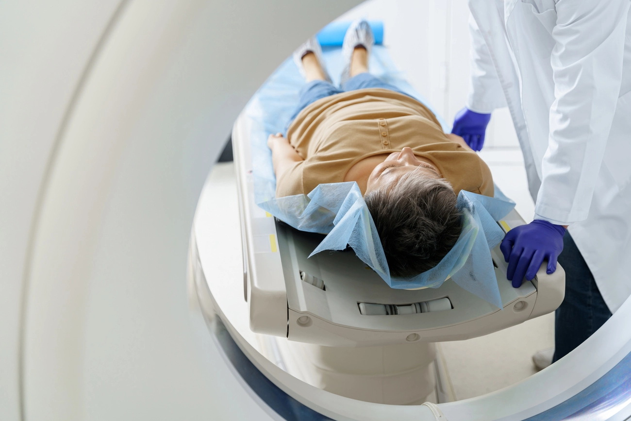
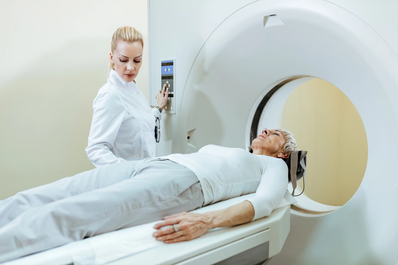
What happens during the brain MRI study?
Here’s a step-by-step guide to what you can expect:
- You will have to remove all the metallic accessories you have on your body.
- Positioning: The technologist will place a helmet-like device called a coil around your head. The brain coil helps the technologist obtain very detailed images of your brain.
- Scanning: You’ll lie on a table that slides into the MRI scanner. You may hear humming or tapping sounds, which are normal. The technologist operates the machine from outside the room and can communicate with you via intercom.
- Duration: The procedure takes 30–60 minutes, depending on the type and complexity of the scan.
- Completion: Once completed, you can return to your daily activities without restrictions.
Results of a brain MRI study: When and how do I receive them?
Brain MRI results are interpreted by a radiologist who sends a detailed report to your doctor.
- Turnaround time: Receive your results in 24 to 72 hours.
- Detailed report: Includes observations of abnormalities and recommendations for further tests or treatments if necessary.
- Follow-up: Your doctor will review the results and determine the next steps.
Benefits of a brain magnetic resonance imaging study
Brain MRIs are invaluable for assessing your brain health. Key benefits include:
- Early detection: Identifies conditions at their earliest stages.
- Accurate diagnosis: High resolution provides reliable results.
- Non-invasive and safe: Unlike other imaging studies, it doesn’t use radiation.
These advantages make brain MRI an essential tool in preventing and treating diseases.

Alternatives to a brain magnetic resonance imaging study
While MRI is highly effective, other imaging options may be considered:
- CT Scan: Faster and cheaper but offers lower resolution for soft tissues and uses radiation.
Conclusion
Brain MRI is a critical study in modern medicine, offering precise and safe diagnostics for various conditions.
This tool is invaluable for brain health, from detecting tumors to monitoring neurodegenerative diseases.
Want to learn more about other diagnostic options? Visit our website to explore studies that can improve your quality of life.